
BLOOD - Light Microscopy
Erythrocytes - Platelets
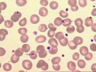 Erythrocytes and platelets as they appear in blood smears stained with
Giemsa.
Erythrocytes and platelets as they appear in blood smears stained with
Giemsa.
Neutrophil - Erythrocytes
 ..
.
..
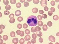
. Images from blood smears revealing the characteristic polymorphic
aspect from the neutrophil nucleus.
Images from blood smears revealing the characteristic polymorphic
aspect from the neutrophil nucleus.
Eosinophil - Erythrocytes - Platelets
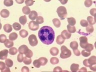
 Blood
smears showing the bilobed nucleus and eosinophilic granules from eosinophil
leukocyte.
Blood
smears showing the bilobed nucleus and eosinophilic granules from eosinophil
leukocyte.
Basophil - Erythrocytes
- Platelets
Image showing 2 basophils. Both the nuclei and
specific granules are stained in purple.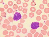
Lymphocyte - Erythrocytes - Platelets
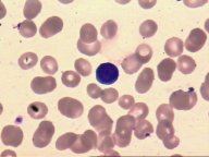 Blood smear showing a lymphocyte between erythrocytes and platelets.
Monocytes - Erythrocytes - Platelets
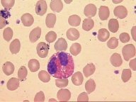
Blood smears with a monocyte, which is bigger
than the surrounding erythrocytes.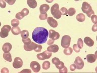
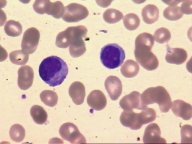 Image showing in the same time a monocyte and a lymphocyte. Despite the
resemblance, it is possible to see that the lymphocyte is a smaller cell
with the nucleus occupying almost all the intracellular space, and the
monocyte, on the other hand, is a bigger cell with a reniform nucleus .
Image showing in the same time a monocyte and a lymphocyte. Despite the
resemblance, it is possible to see that the lymphocyte is a smaller cell
with the nucleus occupying almost all the intracellular space, and the
monocyte, on the other hand, is a bigger cell with a reniform nucleus .