
DIGESTIVE SYSTEM- Light microscopy
Salivary glands, Liver, Gallbladder and Pancreas
Salivary Glands
Sublingual Gland
 Low magnification image from the sublingual gland showing its mucous acini.
Higher magnification image from the gland showing
their acini and ducts.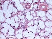
 Detailed image from the mucous acini.
Higher magnification from the anterior image showing
mucous cells from the sublingual gland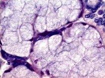
Submandibular Gland
Low mag image from this mixed gland showing mucous,
serous and mixed acini.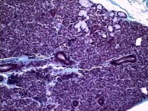
 Higher magnification from the anterior image showing the different types
of acini.
Higher magnification from the anterior image showing the different types
of acini.
In this image it is possible to see the serous,
mucous and mixed acini..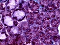
Parotid Gland
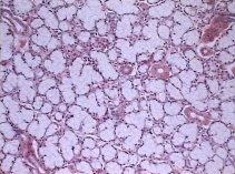
 Low mag image from the gland showing their serous acini. Comparing to the
Pancreas this gland presents a higher amount of connective tissue and adipose
cells.
Low mag image from the gland showing their serous acini. Comparing to the
Pancreas this gland presents a higher amount of connective tissue and adipose
cells.
A higher magnification image from the parotid
showing their serous acini..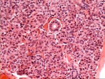
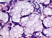
 In
this image it is possible to see the serous acini and intralobular duct
from the parotid gland.
In
this image it is possible to see the serous acini and intralobular duct
from the parotid gland.
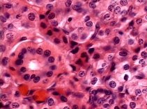
Liver
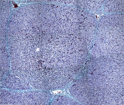 Low mag montage from a slide showing the lobular organization of the liver.
Image showing the portal area located at the lobules
corners. 
Gallbladder
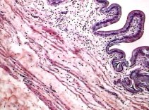 Low mag image from the Gallbladder.
The mucosa from the gallbladder formed by simple
columnar epithelium and lamina propria.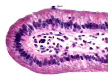
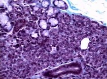
Pâncreas
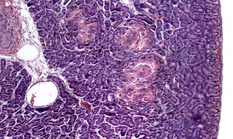
Low mag montage from the pancreas
showing its exocrine and endocrine portions.