
MUSCLE - Electron Microscopy
Transmission Electron Microscopy
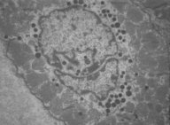
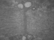
Cardiac Muscle
Central Nuclei
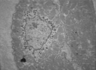 ..
.. Cardiac
muscle in cross section
Cardiac
muscle in cross section
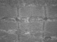
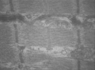
Muscle fibers in longitudinal section
 ..
.. ..
..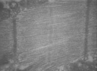
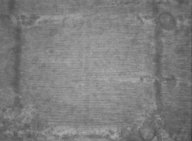
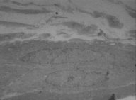
Detail from the striated cardiac fibers in longitudinal
section, revealing the sarcomere organization.
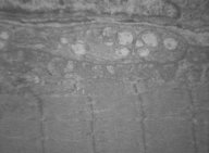
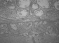
Muscle fibers in cross section
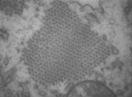 ..
..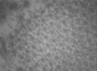 Detail
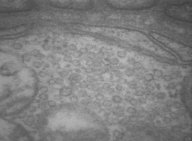
from both actin and myosin organization, seen in cross section.
Detail
from both actin and myosin organization, seen in cross section.
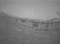
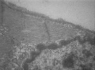
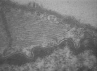
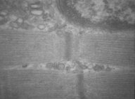
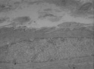
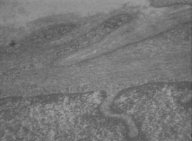
Intercalated discs
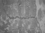 Image showing the connection point between the 2 cardiac fibers, structure
known as intercalated disc, rich in desmosomes.
Image showing the connection point between the 2 cardiac fibers, structure
known as intercalated disc, rich in desmosomes.
Skeletal Muscle
Peripheral Nuclei
 ..
.. ..
.. Several
images showing the peripheral nuclei from the skeletal muscle fibers.
Several
images showing the peripheral nuclei from the skeletal muscle fibers.
Muscle fibers in longitudinal section and triads
 ..
.. ...
... Different
images from the sarcomere in the skeletal muscle and the presence of triads
between the fibrils.
Different
images from the sarcomere in the skeletal muscle and the presence of triads
between the fibrils.
Motor end plate
 ..
.. ...
... Myoneural
junction, where it is possible to see the axon terminal filled with synaptic
vesicles.
Myoneural
junction, where it is possible to see the axon terminal filled with synaptic
vesicles.
Smooth Muscle
 ..
.. ..
.. Smooth
muscle fibers in different magnifications, where it is visible the central
nucleus and the absence of sarcomeric structure.
Smooth
muscle fibers in different magnifications, where it is visible the central
nucleus and the absence of sarcomeric structure.