
NERVOUS TISSUE - Eletron Microscopy
TransmissionEletron Microscopy
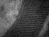 Section of a nerve fiber revealing the myelin shealth.
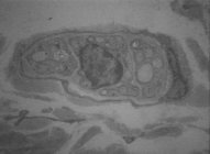
This image shows a synapsis between two nerve
processes. 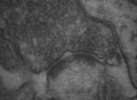
 This
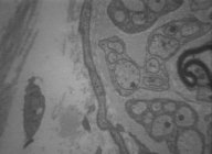
image shows a nerve with myelinated and unmyelinated fibers.
This
image shows a nerve with myelinated and unmyelinated fibers.
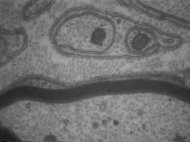 A
higher magnification of the above image showing the myelinated and the
unmyelinated fibers.
A
higher magnification of the above image showing the myelinated and the
unmyelinated fibers.
Space rich in collagen fibers betwenn the nerve
fibers.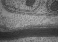
Image showing the epithelioid perfineural cell
with several endocytic vesicles.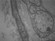
 Image
showing a unmylienated fiber and also the nuclei of Schwann cell.
Image
showing a unmylienated fiber and also the nuclei of Schwann cell.