
NERVOUS TISSUE - Light microscopy
Cerebral Cortex
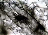 Higher
magnification of the cerebral cortex showing neurons and their processes.
Higher
magnification of the cerebral cortex showing neurons and their processes.
Spinal cord
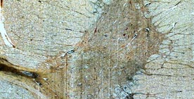 This
image shows the ventral horn of the spinal cord with the distribution of
its dark and white matter.
This
image shows the ventral horn of the spinal cord with the distribution of
its dark and white matter.
Aspect of the dark matter of the spinal
cord, with neurons and processes. 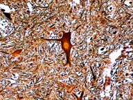
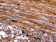 This image shows several neuronal fibers in the
spinal cord white matter.
This image shows several neuronal fibers in the
spinal cord white matter.
Cerebellum
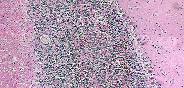 Image
showing the cerebellar layers.
Image
showing the cerebellar layers.
Image of the intersection region between
molecular layer, Purkinje cell layer and granule cell layer of the cerebellum.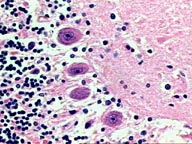
Aspect of the Purkinje cell layer.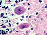
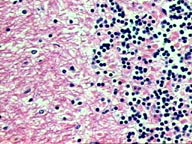 Intersection
region between granule cell layer and the white matter.
Intersection
region between granule cell layer and the white matter.