URINARY SYSTEM - Light Microscopy
KIDNEY
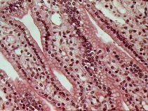
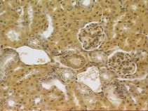 Image from the renal cortex showing the convoluted tubules and renal corpuscules.
Higher magnification from the cortex area showing
one corpuscule and convoluted tubules
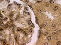 Detailed image from the corpuscule showing the Bowman´s capsule and
the glomerulus.
Detailed image from the corpuscule showing the Bowman´s capsule and
the glomerulus.
Image showing the vascular pole from the renal
corpuscule. 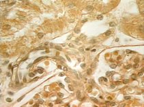
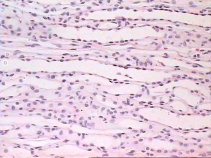 Image from the medullar region of the kidney with some loops of Henle.
Transverse section through the medulla, showing
portions of loops of Henle. 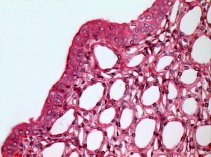 .
.
 Collecting tubules in longitudinal section.
URETER
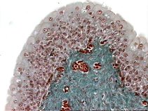
Low mag transverse section from the ureter, showing
the mucosa thrown up into folds..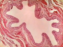
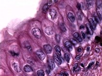
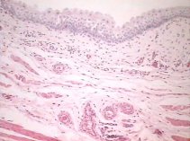 Higher mag image from the ureter, showing the transitional epithelium.
Bladder
 ...
... Images showing the mucosa of the bladder.
Images from the transitional epithelium of the
bladder 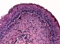 .
.