
Cell - Electron Microscopy
Cells and organelles
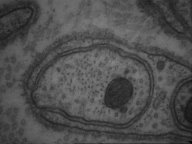 Unmyelinated
nerve fibers surrounded by one layer of myelin from the Schwann cell.
Unmyelinated
nerve fibers surrounded by one layer of myelin from the Schwann cell.
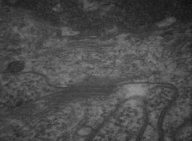
Inner aspect of a cell showing a nucleus, Golgi
complex and a centrioles. 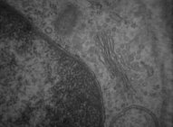
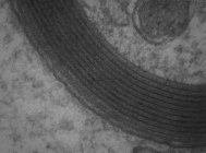 Myelin
sheath originated by a multilayered spiral wrapping of Schwann cell membrane
Myelin
sheath originated by a multilayered spiral wrapping of Schwann cell membrane
Mitochondria and synaptic vesicles in a axonal
termination. 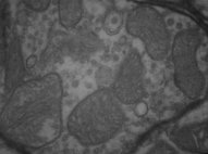
 ..
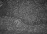
..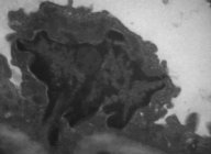 Lymphocyte adhered to a endothelial cell wall.
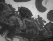
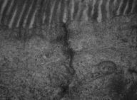
Specializations of the apical surface
Cilia
Transmission Electron Microscopy
 ..
..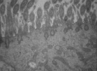 Image showing the apical aspect from ciliated columnar tracheal cells.
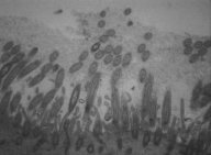
Scanning Electron Microscopy
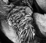 Image
showing the cilia and microvilli from the trachea epithelium.
Image
showing the cilia and microvilli from the trachea epithelium.
Microvilli
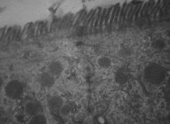
Scanning Electron Microscopy
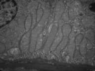
Scanning electron microscopy aspect from the
brush border in the intestinal epithelium. 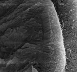
Transmission Electron Microscopy
 ...
...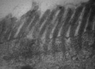 Microvilli
in the apical surface from the intestinal cells.
Microvilli
in the apical surface from the intestinal cells.
Specializations from the lateral membrane - Cells junctions
Junctional Complex
 Junctional
complex between two adjacent intestinal cells.
Junctional
complex between two adjacent intestinal cells.
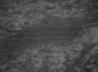
Tight Junction
 Tight junction between two endothelial cells from a capillary. This image
also show the basal lamina and pinocytic vesicles.
Tight junction between two endothelial cells from a capillary. This image
also show the basal lamina and pinocytic vesicles.
Adhesion Junctions and Desmosome
Adhesion junctions and desmosomes from the intercalated
disc in the cardiac muscle.
 ..
..  Desmosome
in the epidermis.
Desmosome
in the epidermis.
Specializations from the basal membrane
Hemidesmosomes
 ...
... Hemidesmosomes
ancoring the keratinocytes from the skin to the underlying basal laminal.
Hemidesmosomes
ancoring the keratinocytes from the skin to the underlying basal laminal.
Branches and convolutions
from the basal membrane
 Branches
in the basal membrane from the kidney convoluted tubule cell.
Branches
in the basal membrane from the kidney convoluted tubule cell.
![]()
![]()