
Cells - Light Microscopy
Cells observed under the light microscopy
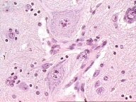
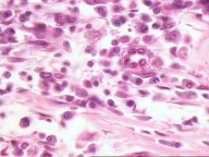 Lymphocytes
and Plasma cells, immune system cells stained by Hematoxilin & Eosin.
Lymphocytes
and Plasma cells, immune system cells stained by Hematoxilin & Eosin.
Endothelial cells from small vessels in the connective
tissue..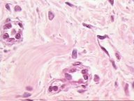
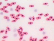 Nucleated
erythrocytes from the frog blood. It is also shown some leukocytes.
Nucleated
erythrocytes from the frog blood. It is also shown some leukocytes.
Stellate mesenchymal cells from a embryonary
tissue .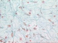
 Neurons
and glial cells from the spinal cord
Neurons
and glial cells from the spinal cord
Purkinje cells, giant neurons from the cerebellar
cortex.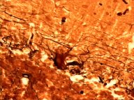
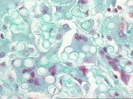 Mutlinucleated giant macrophages from the connective tissue.
Specializations of the apical surface
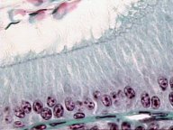
Cilia
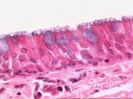 Image
from the ciliated pseudostratified columnar epithelium from the trachea.
Image
from the ciliated pseudostratified columnar epithelium from the trachea.
Microvilli
Image showing the brush border from the absortive
columnar epithelia 
Stereocilia
 Pseudostratified columnar epithelium with stereocilia from the epididymus.
![]()
![]()