
METHODOLOGY and EQUIPMENT (part II)
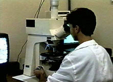 After stainning the slides can be observed in the light microscope.
This model is a Zeiss Axiophot equipped with planapochromatic objectives
and zooming lens before the video-camera.
After stainning the slides can be observed in the light microscope.
This model is a Zeiss Axiophot equipped with planapochromatic objectives
and zooming lens before the video-camera.
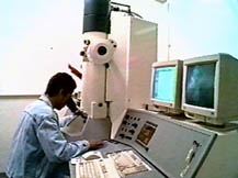 The grids prepared before can now be analysed in the TEM. This model is
a LEO 906, with associated image processing system .
The grids prepared before can now be analysed in the TEM. This model is
a LEO 906, with associated image processing system .
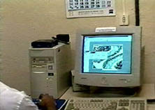 All the microscopes used in the preparation of this ATLAS were connected
to video-cameras. The video sequences generated were recorded in analog
tapes or digitized. In order to create digital files, a computer should
receive the signal from the camera and transform the analog input to
digital data. To perform this job we used PCs equipped with frame
grabbers, that create static images in TIF format or digital films in AVI.
All the microscopes used in the preparation of this ATLAS were connected
to video-cameras. The video sequences generated were recorded in analog
tapes or digitized. In order to create digital files, a computer should
receive the signal from the camera and transform the analog input to
digital data. To perform this job we used PCs equipped with frame
grabbers, that create static images in TIF format or digital films in AVI.
 In order to present the images and make easier its transmission by the
internet, the TIF files were converted to a compact form, JPEG. This means
that some resolution were lost during compression proccess. This decrease
in resolution is more visible in the TEM images. Another important
tool used was the computer montage of several frames to generate a high
resolution and wide image field. These images are always presented
in the initial page of each tissue and system in the ATLAS.
In order to present the images and make easier its transmission by the
internet, the TIF files were converted to a compact form, JPEG. This means
that some resolution were lost during compression proccess. This decrease
in resolution is more visible in the TEM images. Another important
tool used was the computer montage of several frames to generate a high
resolution and wide image field. These images are always presented
in the initial page of each tissue and system in the ATLAS.