
 All the material used in this ATLAS, independent of the microscope used
to visualized had to be fixed shortly after its removal. The process of
fixation changes a little from light to electron microscope. In general
the material analyzed in light microscopy was fixed in a solution containing
formaldehyde 10% in buffer and the samples observed in the electron microscope
were fixed in a solution containing 2.5% of gluteraldehyde and 4% of paraformoldehyde
in buffer. All material were kept in 40 Celsius.
All the material used in this ATLAS, independent of the microscope used
to visualized had to be fixed shortly after its removal. The process of
fixation changes a little from light to electron microscope. In general
the material analyzed in light microscopy was fixed in a solution containing
formaldehyde 10% in buffer and the samples observed in the electron microscope
were fixed in a solution containing 2.5% of gluteraldehyde and 4% of paraformoldehyde
in buffer. All material were kept in 40 Celsius.
 After
the fixation, the samples were rinsed several times, dehydrated and included
in resin, (the type of resin used also varies in function of the microscope
used, light or transmission electron) the material must be
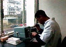
sectioned. In light microscopy, we use a equipment called Microtome, to
obtain sections with thickness of few micrometers (1mm
= 10-6 m).
After
the fixation, the samples were rinsed several times, dehydrated and included
in resin, (the type of resin used also varies in function of the microscope
used, light or transmission electron) the material must be
sectioned. In light microscopy, we use a equipment called Microtome, to
obtain sections with thickness of few micrometers (1mm
= 10-6 m).
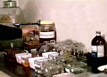 The
sections were collected using a slide, and after few steps the samples
were stained with different types of stainning solutions. The most famous
stainning solution is the Hematoxiline & Eosin. This solution called
by H&E stains the nucleus in purple and the cytoplasm in rose.
The
sections were collected using a slide, and after few steps the samples
were stained with different types of stainning solutions. The most famous
stainning solution is the Hematoxiline & Eosin. This solution called
by H&E stains the nucleus in purple and the cytoplasm in rose.
After drying, the slides must
be mounted with a coverslip. To do so, a drop of mounting oil is
settle over the material and the coverslip is sealed. The slide is now
ready to be observed in the light microscope.
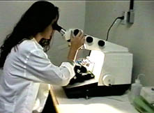 The
samples that will be observed in the transmission electron microscope (TEM)
must also be sectionned. This is performed in an equipment called Ultramicrotome.
The sections obtained in this equipment are in the range of 40 - 60 nanometers
(1nm= 10-3mm), in order to be crossed
by the electron beam. The sections were collected with very small
grids and as happened with the light microscope, must be stainned previously
to the observation. To perform that we use solutions containing
metals with high atomic number, such as lead, uranium and osmius.
The
samples that will be observed in the transmission electron microscope (TEM)
must also be sectionned. This is performed in an equipment called Ultramicrotome.
The sections obtained in this equipment are in the range of 40 - 60 nanometers
(1nm= 10-3mm), in order to be crossed
by the electron beam. The sections were collected with very small
grids and as happened with the light microscope, must be stainned previously
to the observation. To perform that we use solutions containing
metals with high atomic number, such as lead, uranium and osmius.